Blog

Weekly Reflection #9

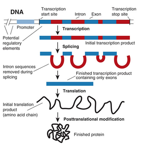
This week in AP Bio we studied gene expression and central dogma. We did a bunch of packets and lectures throughout the week to introduce it. First, we looked into central dogma – the flow of genetic information from DNA to proteins, then DNA coding to RNA, then DNA to RNA to proteins. There are two main steps – transcription and translation. First is transcription which is where DNA is being transcribed into mRNA which can be divided up into four stages.

  • Initiation
    • The DNA molecule unwinds and separates and creates small opening where RNA polymerase binds to the promoter of the template DNA strand. In prokaryotes RNA polymerase binds directly to the promoter and in Eukaryotes RNA polymerase requires an assemblage of transcription factor proteins to be able to bind to the promoter.
  • Elongation
    • Moves in a 5′ to 3′ direction. RNA polymerase goes along the template strand, creating a mRNA strand containing the important genetic information. The coding strand will have the same sequence with thymines replaced by uracil.
  • Termination.
    • In prokaryotes there are two ways in which transcription is terminated. Rho-dependent termination- the Rho protein destabilizes the RNA-DNA hydrogen bonding at RNA polymerase and ceases transcription. In Rho-independent termination, the transcript bases hydrogen bond with themselves, fold back and pull the transcript out of the RNA polymerase.
  • Post-transcriptional mRNA Processing
    • Introns are removed and the exons are spliced together to form a mRNA molecule consisting of a single protein-coding sequence.

Image result for transcription of dna

Next is translation, which is made up of a similar but different process involving 3 steps; initiation, elongation and termination.

  • Initiation
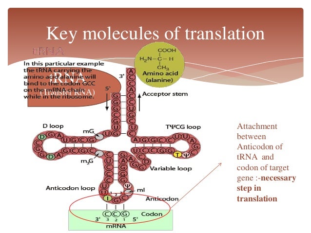
    • The mRNA attaches to the small ribosomal subunit o at the 5′ end of the mRNA molecule and moves in a 3′ direction until it meets a start codon (AUG) at the P-site. tRNA binding at the ribosome is mediated by an “anti-codon” loop in the tRNA molecule.
  • Elongation
    • The ribosome travels down the message, reading codons and bringing in the proper aminoacyl tRNA’s to translate the message to the protein.  The new tRNA is brought into the ribosome A site, where it is matched with the codon being presented.
  • Termination
    • Translation is stopped when a stop codon (UAA, UAG, UGA) is encountered and a release factor binds to the A-site. The polypeptide chain is released and the ribosome disassembles.

Image result for translation of dna

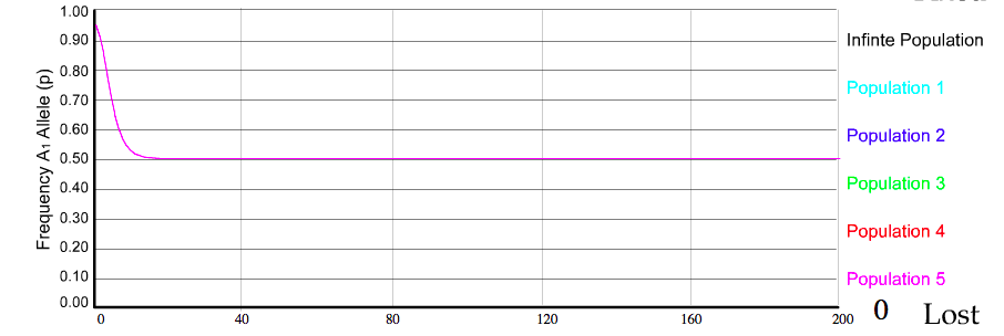
Next, we can take look at gene expression. There are two types of gene expression which include inducible (it turns the operon on, starts transcription and translations, caused by a new metabolite which needs enzymes to get metabolized, operates in a catabolic pathway, and the repressor is prevented by the inducer from joining the operator gene) and repressible (it turns the operon off, stops transcription and translation, caused by an excess of existing metabolite, operates in an anabolic pathway, and aporepressor is enabled by a co-repressor to join the operator gene).

Image result for gene expression

This week was focused on the big idea 2 – understanding within living organisms. I enjoyed doing the packet regarding dog genes this week as it really stimulated my thinking process and made me work for it in an entertaining way vs. the other packets when you have to google to find the answers. Next week I want to look more at diseases in this area, and how translation/transcription can go wrong in organisms.

Websites

Weekly Reflection #10

This week in AP Biology, we did an intense lab for most of the week after a quick test on Monday. The lab was long and strenuous, but taught us what happens to your body when you inhale and exhale.

In the introduction of the lab, we get a quick background to photosynthesis and cellular respiration. Along the way, we answer “Thinq Exercises” to expand our knowledge and stimulate our brains. A lot of the questions were random and irrelevant but whatever floats your boat Bio-Rad. The lab goes into depth about interdependent pathways – for example, how cellular respiration relies on photosynthesis. Then we answered a few more questions about the Hill Reaction and how it powers the Calvin Cycle.

Starting investigation #1, we found ways to monitor photosynthesis AND cellular respiration by using the same system. We also looked a bit into examples of autotroph producers in the real world, and how they are able to live in their environments. Next, we identified how one process can be identified over another. In the light, algae can perform both photosynthesis and cellular respiration but in the dark, it can only perform cellular respiration.

We performed the Algae Microscopy lab by depolymerizing the algae beads to free the algae and study what it looked like under a microscope. We came to see that some S.obliquus were varying in color.

In investigation 2 (the longest part), we used a CO2 indicator to show when CO2 levels increase or decrease during photosynthesis and cellular respiration. We transferred algae beads to different substances and placed them under light, carefully monitoring the color and pH changes to identify what was happening.

This was a long week with a long lab, but its better than taking notes! Hopefully next week we can look into labs that are a little more active vs. waiting around for an hour to see what color the algae beads change to.

WEBSITES

https://study.com/academy/lesson/the-relationship-between-photosynthesis-cellular-respiration.html

https://prezi.com/nxdgg-kutyji/interdependence-of-cellular-respiration-and-photosynthesis/


Weekly Reflection – 10

This week in AP bio, we progressed from the cellular respiration unit to more specifically, the photosynthesis unit. On Monday we resumed one more lecture to finalize the cellular respiration unit, then on Tuesday we started lecturing on photosynthesis. We did a few online packets to help us gain basic knowledge first as well.

On Monday, we started with learning that most cellular respiration requires O2 to produce ATP and without O2, the electron transport chain will cease to operate. In that case, glycolysis couples w fermentation or anaerobic respiration to produce ATP. The formula for cellular respiration is C6H12O6 + 6O2 → 6H2O + 6CO2. 

Anaerobic respiration uses an electron transport chain w a final electron acceptor other than O2 for example sulphate. Fermentation uses substrate level phosphorylation instead of an electron transport chain to generate ATP. Fermentation consists of glycolysis plus reactions that regenerate NAD+, which can be reused by glycolysis afterwards. Two common types are alcohol fermentation and lactic acid fermentation.  In alcohol fermentation, pyruvate is converted to ethanol in two steps: releasing CO2 from pyruvate, then reducing acetaldehyde to ethanol. An example of alcohol fermentation being used in a real life scenario is yeast being used in brewing, winemaking and baking. 

Lactic acid fermentation is a completely different type though. Pyruvate is reduced by NADH, forming lactate as an end product w no release of CO2. An example of lactic acid fermentation in a real world scenario is when some fungi and bacteria is used to make cheese and yogurt. Human muscle cells also use lactic acid fermentation to generate ATP when O2 is scarce.

In comparing fermentation with anaerobic vs aerobic, we must acknowledge a few things. All use glycolysis to oxidize glucose and harvest chemical energy of food and in all 3, NAD+ is the oxidizing agent that accepts electrons during glycolysis. The processes have different final electron acceptors: an organic molecule (pyruvate) in fermentation and O2 in cellular respiration. Cellular respiration produces 32 ATP per glucose mol: fermentation produces 2ATP per glucose mol. Obligate anaerobes carry out only fermentation or anaerobic respiration and cant survive in the presence of O2. Yeast and many bacteria are facultative anaerobes, meaning that they can survive using either fermentation or cellular respiration. In a facultative anaerobe, pyruvate is a fork in the metabolic road that leads to two alternative catabolic routes.

FINALLY we can get into photosynthesis. We delved into what actually happens during photosynthesis and what drives the reaction.

I learned that CO2 comes into  stoma, oxygen and water come out. Water needs to evaporate to drive the process of taking water up into the leaves – creates negative pressure so it fills void. NADPH is created by photosystem I. The main ingredient is carbon dioxide – binds to RuBP. To combine them, we use an enzyme called RuBisCo. It takes CO2 and sticks it to 5 carbon sugar to create a 6 carbon sugar (becomes unstable) so then the sugar breaks down. C3 plants do great in cool weather but suffer in cold weather – by why? It’s because the CO2 concentration is used up throughout the day while oxygen increases because there’s no way out. Therefore RuBisCo is more likely to bind w oxygen, so it takes components out of the reaction that should function properly – this is also known as photorespiration. It prevents current photosynthesis by taking reactants out of the cycle. We also addressed questions such as “How can we have plants in a hot location?” The answer is to have an open stomata at night so less water will evaporate. It will store CO2 at night to be used for reactions during the day OR you can do the light dependent reactions somewhere else so O2 won’t join to RuBisCo. The xylem is responsible for moving water soluble things from the ground to the plant while the phloem is responsible for taking the products and moving it to where it needs to go.

This week we did more lecturing than I’d like to, but I think I got a good grasp on the basics of photosynthesis. As for the labs, I sped through the virtual ones relatively easily. I’m excited to see what we’ll be doing for the rest of the photosynthesis unit, hopefully do some labs involving plants.

WEBSITES

http://www.phschool.com/science/biology_place/biocoach/cellresp/intro.html

https://www.khanacademy.org/science/biology/cellular-respiration-and-fermentation/variations-on-cellular-respiration/a/fermentation-and-anaerobic-respiration

https://www2.estrellamountain.edu/faculty/farabee/BIOBK/BioBookPS.html


Weekly Reflection – 9

This week in AP Biology we focused cellular respiration. Cellular respiration is the process by which the chemical energy of “food” molecules is released and partially captured in the form of ATP. Carbohydrates, fats, and proteins can all be used as fuels in cellular respiration, but glucose is most commonly used as an example to examine the reactions and pathways involved.

We can divide cellular respiration into three metabolic processes: glycolysis, the Krebs cycle, and oxidative phosphorylation. Each of these occurs in a specific region of the cell.

  1. Glycolysis occurs in the cytosol.
  2. The Krebs cycle takes place in the matrix of the mitochondria.
  3. Oxidative phosphorylation via the electon transport chain is carried out on the inner mitochondrial membrane.

In the absence of oxygen, respiration consists of two metabolic pathways: glycolysis and fermentation. Both of these occur in the cytosol. In glycolysis, the 6-carbon sugar, glucose, is broken down into two molecules of a 3-carbon molecule called pyruvate. This change is accompanied by a net gain of 2 ATP molecules and 2 NADH molecules.

The citric acid cycle occurs in the mitochondrial matrix and generates a pool of chemical energy (ATP, NADH, and FADH2) from the oxidation of pyruvate, the end product of glycolysis. Pyruvate is transported into the mitochondria and loses carbon dioxide to form acetyl-CoA, a 2-carbon molecule. When acetyl-CoA is oxidized to carbon dioxide in the citric acid cycle, chemical energy is released and captured in the form of NADH, FADH2, and ATP. The electron transport chain allows the release of the large amount of chemical energy stored in reduced NAD+(NADH) and reduced FAD (FADH2). The energy released is captured in the form of ATP (3 ATP per NADH and 2 ATP per FADH2). The electron transport chain (ETC) consists of a series of molecules, mostly proteins, embedded in the inner mitochondrial membrane. All cells are able to synthesize ATP via the process of glycolysis. In many cells, if oxygen is not present, pyruvate is metabolized in a process called fermentation.

There are two different kinds of fermentation. The first is lactic acid fermentation, this is when pyruvate is reduced by NADH and lactase is formed as the product. The other kinds is alcohol fermentation, which is when pyruvate is converted into ethanol, releasing CO2, and oxidizing NADH which produces more NAD+.

Glycolysis is another main part of cellular respiration,  chemical energy is harvested by oxidizing glucose into a pyruvate. This is also known as “sugar splitting”. It occurs in two major phases: the energy investment phase and the energy payoff phase. Pyruvate is oxidized, the citric acid cycle completes the enrgy-yielding oxidation of organic molecules. The chemical energy stored in glucose generates far more ATP in aerobic respiration than in respiration without oxygen (glycolysis and fermentation). Each molecule of glucose can generate 36-38 molecules of ATP in aerobic respiration but only 2 ATP molecules in respiration without oxygen (through glycolysis and fermentation).

Chemiosmosis is the energy coupling mechanism. The electron transport chain pumps H+ ons into mitochondria membrane . Energy in the hydrogen ion gradient to drive cellular works. This happens oxidative phosphorylation, where it couples with the electron transport chain to synthesis ATP.

We also did a quick lab on the last day that involved us deducing the disease by looking at symptoms of several different victims. We did this by analyzing the death of their mitochondrial cells.

I would love to do more labs similar to the one above, where you diagnose patients and put your knowledge to the test.

Websites Used:

https://www.scq.ubc.ca/protein-phosphorylation-a-global-regulator-of-cellular-activity/

https://www.khanacademy.org/science/biology/cellular-respiration-and-fermentation


Reflective Response Week 8

This week in AP Biology we focused on the structure and support mechanisms of the cell.  The organized structures within the cell are called organelles – each one is specific in helping the cell to function as a working group.

The first organelle we looked into was the cytoskeleton. The cytoskeleton is a network of structural proteins that are connected in the cytoplasm. Microtubules are found in the cytoskeleton and are made up of tubulin monomers which are used as the transportation system for vesicles. Microfilaments and intermediate filaments are also found in the cytoskeleton, which are both made up of actin monomers and help with cell division and protection of the nucleus. The cytoskeletons function is to help with the movement, protection, and structure of a the cell.

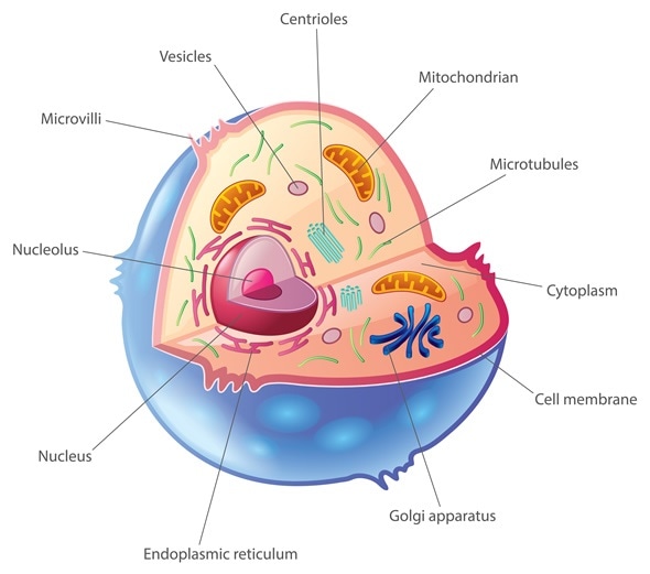
The cell membrane is made up of a phospholipid bilayer with proteins, otherwise known as the “Fluid mosaic model”. This is like the security gate into the cell, helping with boundary, transportation, and communication. The phospholipid found in the membrane is amphipathic, meaning it is both polar and non polar, and create a semi-permeable membrane. The membrane proteins include both integral and peripheral proteins. Integral penetrate both bilayers, while peripheral proteins don’t penetrate but are hovering on top. These assist in creating polarity. The Cilia and Flagella are motility related extensions of cytoskeletal proteins. The centrosome is the microtubule and is only found in animal cells.

Cholesterol is a steroid lipid that act as a “temperature buffer” to help with membrane fluidity. Integral proteins span the bi-layer with domains and function with signal translation. Glycoproteins also span the bi-layer but they have short polysaccarides vesicles projecting which serve as identifying maker in cellular populations. Cell walls, on the other hand, are only contained within plant cells. These are too rigid to withstand the turgid pressure for animal cells, so in place we use an extracellular matrix. The “ECM” is a network of connective proteins and protoglycen molecules outside of the cell membrane. This assists in the cell anchorage and communication.

This week we learned much more about the specific functions of organelles, which was useful in figuring out more of the cellular purpose. I’m excited to learn about more organelles such as the mitochondria and Golgi apparatus!

Websites:

Learn About Organelles – ThoughtCo

Cell Membranes | Learn Science at Scitable – Nature


Reflection Week 7

This week in AP Biology we started to talk about cell signaling and membrane transport, which ties in with big question #2. First, we talked about diffusion. Diffusion is the spreading of something from a higher concentration to a lower concentration. On Tuesday, we did a lab that had visual examples of the ways cells spread. The main goal of molecules in diffusion is to take up the space given in an equal distribution.

Next, we talked about the main two types of transport which are:

Active: Active transport does require energy. It uses protein and enzymes to control what is coming in and out of the cell. If there were to be a specific protein for a molecule that would make it change to facilitated diffusion.

Passive: Passive transport doesn’t require energy because it follows the natural process of molecule movement from a high concentration to a low one. It is based of the random movement of molecules. Channels are carrier proteins control the diffusion of charged or polar molecules.

Tonicity is the measurement of relative measure of a solution concentration. We learned about three different types of cell solutions.

Hypotonic solution: Where there is less solution in cell than out, so solution wants to diffuse till at equilibrium. These cells can pop.

Hypertonic solution: Basically the opposite of a hypotonic solution, more solution inside the cell than outside, so wants to diffuse till equal. These cells can shrivel up.

Isotonic solutions- equal amounts of solution inside and outside cell. Ideal situation.

Below is a visual representation of hypertonic, hypotonic and isotonic solutions.

Unknown.jpeg

There are two types of bulk transport that help bring out or kick out large amounts of molecules.

Endocytosis: the taking in of matter by a living cell by invagination of its membrane to form a vacuole. Phagocytosis: “cell eating”, Pinocytosis: ” cell drinking”, Receptor- mediated endocytosis: “cell being picky” are all subtypes of endocytosis.

Exocytosis: the release of cellular products into outflow

As far as labs go, we did an electronic one on Tuesday that helped us understand the basic mechanisms of cell transport and one on diffusion and osmosis on Thursday. These both extended my knowledge of biochemistry and solidified many of the basics for me.

Next week I would like to learn more about the purpose of different types of molecules going in and out of the body and why they do what they do.

Websites:

https://www.khanacademy.org/test-prep/mcat/cells/transport-across-a-cell-membrane/a/passive-transport-and-active-transport-across-a-cell-membrane-article

https://www.diffen.com/difference/Diffusion_vs_Osmosis

Reflection Week 6

This week in AP Biology, we started off with learning a little bit about water’s properties and specific heat, then delved deeper into the complicated world of proteins. Proteins are responsible for all life activities of the cell.

Water is special and is considered most like a “universal solvent” because not only does it exist in all 3 phases (liquid, solid and gas) at normal terrestrial conditions, but its solid form is less dense than its liquid form. This is because the hydrogen bonds are an ideal distance away from each other, as well as having very high surface tension.

Next, we focused on proteins. Proteins are made up of carbon, hydrogen, oxygen, nitrogen, and sometimes sulfur. From these elements, amino acids are created to make up monomers inside of polymers. From what scientists know right now, there are only 21 amino acids. Every amino acid has a different function within the protein due to the different structure of the R group. The directionality of the amino acid is determined from the amino group to the carboxyl group (on opposite ends of the structure). There are 4 different types of structure within the protein.

First, there is the Primary structure. The primary structure is a peptide chain made up of amino acids bonded by peptide bonds, or covalent bonds (the strongest).

Then there is Secondary structure. This includes regular repeated 3D structures found in all polypeptide chains, with hydrogen bonding between atoms in the Carbon-Nitrogen backbone. The two different forms are the alpha helix and beta pleated sheet.

Tertiary structure is the next level up, including a 3D shape of a particular polypeptide chain (conformation). This comes from interactions between the R group atoms with other R group atoms and their environments in the cell.

Quaternary structure is the 3D shape of any protein that is made of more than one polypeptide chain, and is the only optional level of the structures. It is the overall appearance when multiple chains form a functional protein.

When proteins “denature”, it means the conformation of the structure has been altered, so therefore the function of the protein will also be altered.

ENZYMES

After learning about proteins, we discussed enzymes. Enzymes (proteins) are responsible for the reactions within the body. Most reactions occur spontaneously by bumping into each other. This requires a certain amount of activation energy, which is why if the molecules are moving faster, there will be more reactions. If the molecules are moving slower, there will be less. Introducing heat speeds up the system and therefore the reactions.

The energy can be divided into to categories; catabolic and anabolic. Catabolic is when bonds are broken down, while anabolic is when bonds are built.

Each enzyme has an active site that fits a specific substrate – this is known as the lock and key model. “Induced fit” is when the enzyme hugs the substrate tightly. The rate of enzyme reactions with their substrate is dependent on the concentration of the substrate and the enzyme, as well as the temperature. Another factor is the types of inhibitors that can appear; you can have competitive or noncompetitive/allosteric inhibitors that prevent reactions.

LABS

Finally, we did two labs that assisted in our understanding of proteins and enzyme reactions. The first we did was pretty fun, when all we did was create cheese curds from milk and vinegar and poach an egg (ours was perfect). This lab showed us how we can put chemical reactions into real life scenarios. The second lab we did was more relevant, and showed us how the process of substrate and enzyme reactions occurred. We did this in three stages, also looking at what happens when you add in inhibitors.

This week, we studied more of big idea #2, which talks about energy in order to maintain homeostasis. Now that we’ve studied the energy aspect, I am expecting to look more into maintaining homeostasis next week.

Links

Orders of Protein Structures

Enzyme Kinetics

Effects of Inhibitors on Enzyme Activity

Reflection Week 5

This week in AP Biology, we focused primarily on the basics of Molecular Biology so we have a level to learn more complicated things from. We started off the week with a video from the weird world series to dismiss a few of the Monday blues. On Tuesday, we covered a few labs and video worksheets in which we answered the basics of biochemistry. On Thursday, the hardest day in my opinion, we did a lab where we matched molecular structures to their groups and names.

On Tuesday, we discussed the 4 different types of major macromolecules.

  1. Lipids include fats and waxes – they consist of one monomer and they store energy.
  2. Nucleic Acids (ex. DNA, RNA) nucleotide monomers, made of adenine, thymine, guanine, cytosine, and uracil.
  3. Proteins (ex. collagen, hemoglobin) amino acid monomers
  4. Carbohydrates (ex. glucose, sucrose) supply energy, sugars

The types of molecules can be more broadly categorized into:

  1. Monomer: Molecules that can be bonded to other monomers, “building blocks of life”
  2. Polymer: A large molecule or macromolecule composed of repeated subunits

On Thursday, we focused on how to take these molecular properties and use them to divide different macromolecules into groups. These could have been subcategories such as nucleotides, ribose and fatty acids. This lab was incredibly hard for me to mentally wrap my head around it, and I’m still working on it as we speak. Carbon, hydrogen and oxygen make up all parts of molecules, but sometimes nitrogen, sulfur and phosphorus are also present. In amino acids, the molecules are focused around a central carbon backbone and has a double bond with oxygen. Steroids, or cholesterol,  have multiple rings of carbon atoms and the carbon sits invisibly at every angle in the diagram. For this reason the beehive-shaped m=diagrams were usually distinguished as steroids. Fatty acids are the long chained diagrams and are a type of lipid. They’re made of hydrocarbon chains with a carboxyl group at one end. In carbohydrates there is about a 1:1 ratio of oxygen to hydrogen and these sugars can be divided into monosaccharides, disaccharides, and straight chain sugars. With the nucleic acids we observed simple and complex nitrogenous bases. These molecules can have nitrogen in them.

This week was difficult for me to understand since I didn’t have a very good experience with chemistry class, but I really want to learn and improve my knowledge of it. This topic delves into the science behind big idea 2, and the building blocks behind all living organisms. This is to ensure that we grasp the basics, so we can use the knowledge to understand more complicated topics.

In the future, I would love to understand more about how different structures are formed in polymers and how I can identify one macromolecule and differentiate it to others.

Sources:

Sciencing: 4 Macromolecules of Life

Macromolecules for Identification


Week 4 Reflective Response

This week in AP Biology, we started with a brand new lab involving investigating microevolution by using a population generation program. Through this lab we disturbed each of the 5 standards of Hardy Weinberg equilibrium in order to see how the population evolves in response. I decided to experiment with the heterozygote advantage within a population, which revealed their dominant and recessive allelic equilibrium. This lab is using a quantifying manner to approach Big Idea #1 by using our knowledge of the 5 standards of Hardy Weinberg equilibrium to experiment and visualize what is happening within the population.

Screen Shot 2017-10-08 at 4.09.20 PM

In class, we discussed 2 new major ideas: speciation and classification. A species is a population whose members can interbreed and produce viable and fertile offspring. Speciation is created by a series of evolutionary processes that result in the reproductive isolation of a population. Darwin called it the “mystery of mysteries”. There are two types of speciation: Allopatric and Sympatric.

Allopatric is a population isolated due to physical barriers. An example of this would be when the Grand Canyon formed, it separated one species of squirrel. The squirrels evolved on their side and now no longer recognize one another as a mate. An example of allopatric speciation is the separation of marine creatures on either side of Central America when the Isthmus of Panama closed about 3 million years ago, creating a land bridge between North and South America. Nancy Knowlton of the Smithsonian Tropical Research Institute in Panama has been studying this geological event and its effects on populations of snapping shrimp. She and her colleagues found that shrimp on one side of the isthmus appeared almost identical to those on the other side — having once been members of the same population.

On the other hand, sympatric speciation is when a species remains in the same physical area but become reproductively isolated from one another. For example, gene flow from one part of the population of flies stops from the other when they choose to lay their eggs in different locations. These are all ways a species can be isolated from one another genetically (isolationism). However, there can also be a hybrid population which is caused by gene flow between two species. A real life example of this would be when 200 years ago, the ancestors of apple maggot flies laid their eggs only on hawthorns — but today, these flies lay eggs on hawthorns (which are native to America) and domestic apples (which were introduced to America by immigrants and bred). Females generally choose to lay their eggs on the type of fruit they grew up in, and males tend to look for mates on the type of fruit they grew up in. So hawthorn flies generally end up mating with other hawthorn flies and apple flies generally end up mating with other apple flies. This means that gene flow between parts of the population that mate on different types of fruit is reduced.

Secondly, we learned how to classify species using the old school method and the new school method. The old school method was created by Carl Linnaeus based off of the hierarchy of life. This was based on domain and kingdom. There were three domains: Bacteria, Eukarya, and Archaea and five kingdoms: Monera, Protista, Fungi, Plantae, and Animalia. This system proved to be inefficient because there were too many different species that were interrelated to one another and this discounted everything we could not see (microscopic) as one species. The new school method was used DNA to find relativity to other animals. We could now look deeper than the surface value of physical appearance and see the genetic code of the species by using cladograms (which show us how they evolved from one common ancestor).

To create a cladogram, you must:

  1. Determine how many species have a particular characteristic in common
  2. Group the species so that the most number of species have the most characteristics in common
  3. Apply the rules of maximum parsimony and likelihood
  4. Sort the types of groups within a cladogram into monophyletic (all descendants of 1 common ancestor), paraphyletic (some he descendants have 1 common ancestor), or polyphyletic.

This big idea showed us how exactly species evolve, and from who/what original ancestors. I would love to explore further spectrums of speciation and classification by being able to sort our own species and classify them in a program or “game”.

Websites

Sympatric Speciation

Allopatric Speciation – PBS

Week 3 Reflective Response

This week, we learned about the Hardy-Weinberg equation and relating theory. It hypothetically introduces a non-evolving population that preserves allele frequencies in order to serve as a model for comparison. In nature, populations can never be in Hardy-Weinberg equilibrium because of 5 things:

  1. There can be no genetic mutations in the DNA – it would create genetic diversity
  2. No sexual selection, only random selection – otherwise a certain trait will be favored in an environment
  3. No genetic drift – environmental changes can lead to division of population
  4. No migration – can’t be influenced by other environments that could stimulate new traits
  5. No competition – predators won’t kill one trait off, so new traits develop

The Hardy-Weinberg equation is p+q=1 for allele frequencies and p^2+2pq+q^2=1 is for genotype frequencies. The p stands for the dominant allele, or p^2 homozygous dominant. The q stands for recessive allele, or q^2 for homozygous recessive. The 2pq stands for the heterozygous representation. The recessive allele (q) is what creates a defective protein, vs. the dominant allele (p) making regular proteins. In order to solve this equation in an example problem, here is what you do:

  • In a population, the dominant phenotype of a certain trait occurs 91% of the time. What is the frequency of the dominant allele?

Since the dominant phenotype shows up in both heterozygous and homozygous dominant genotypes, you subtract .91 from 1. This will give you .09, which is the recessive genotype (q^2). Next, find the square root (.03) which is the recessive allele frequency (q). Take this value away from 1 because p+q=1. You get 7 – square 7 (.49) to find the homozygous dominant genotype frequency (p^2). Then you can take the original values to plus in to 2pq to find the heterozygous genotype frequency (.42). Now that you have all the data, you can determine that the dominant allele frequency is .7 or 7%.

Putting this theory in practice, during class we did a collective lab in which we imitated mating within a population. We attempted creating an ideal Hardy-Weinberg population,  selecting against homozygous recessive, examining the heterozygous advantage and genetic drift in populations. Although our results were similar to what we expected, they weren’t perfect because we aren’t an unlimited population like in nature. Through this, we also realized that it is naturally impossible to achieve all of the five conditions to make a Hardy-Weinberg population.

To conclude, this week we took our knowledge of big idea #1 and learned how to measure it and sort data from it. Although it wasn’t necessarily 100% correct, it still communicated the main ideas of population genetics. I would love to learn about more ways to sort biological data and calculate it through other equations because once I understood how to use the Hardy Weinberg formula, it was fun to use!

Websites:

Hardy Weinberg Formulas – KSU

The Heterozygote Advantage

The Ideal Population